e-Learning • EMERALD - European Medical Radiation Learning Development, training modules in medical radiation physics (X-ray Diagnostic Radiology, Nuclear Medicine, Radiotherapy) • EMIT - European Medical Imaging Technology Training pilot project to develop a work-linked training in hospitals on Ultrasound and Magnetic Resonance Imaging Technology • EMITEL - European Medical Imaging Technology e-Encyclopaedia for Lifelong Learning: the first e-Encyclopaedia of Medical Physics with Multilingual Dictionary of terms
Practical and Theoretical Medical Physics Course Series 1 : Quality Assurance (QA) of Advanced Radiotherapy Date: 25-26 August 2016 Venue: Radiotherapy Unit, Advanced Medical & Dental Institute, Universiti Sains Malaysia, Bertam, Penang Course Fee: RM650.00
Course overview: The course is designed to provide the necessary practical and theoretical background in medical physics. The first course of the series covers the radiotherapy physics service of image guided and intensity modulated radiotherapy (IG-IMRT) focusing on the quality assurance aspect. The sessions include practical aspects on quality assurance practice for IM-IGRT including IGRT image quality and IMRT dosimetry verification. The course is aimed primarily at radiotherapy physicists, but should also benefit postgraduate students, researchers, engineers, radiographers, manufacturer’s representative and anyone interested to update their knowledge and understanding of IG-IMRT. The course is CME accredited by Ministry of Health Malaysia.
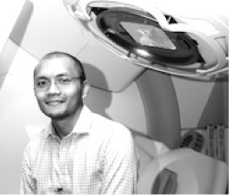
• Qualification: PhD (Radiotherapy Physics), Institute of Cancer Research, London • Research interests: Application of novel image sensor and radiation detector in radiotherapy to improve targeted radiotherapy treatment of cancer using optimised image guidance modalities and real-time MLC tracking • Email: Alamat emel ini dilindungi dari Spambot. Anda perlu hidupkan JavaScript untuk melihatnya. • Phone number: (+6) 04-562 2551 • Website: http://www.amdi.usm.my/oncohafizmz
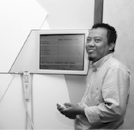
{tab=Dr Mohd Zahri Abdul Aziz}
• Qualification: PhD in Heath Science (Medical Radiation Physics) • Research interests: Monte Carlo Simulations, Dose Calculation Algorithm, Radiation Dosimetry, Electron Beam Therapy, Image Processing • Email: Alamat emel ini dilindungi dari Spambot. Anda perlu hidupkan JavaScript untuk melihatnya. • Phone number: (+6) 04-562 2356 • Website: http://www.amdi.usm.my/oncozahri
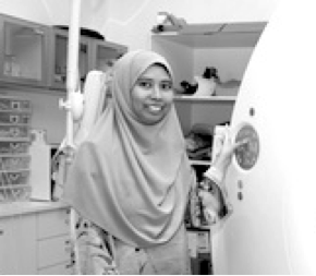
{tab=Dr Noor Diyana Osman}
• Qualification: PhD in Medical Radiation Physics, UiTM • Research interests: Medical physics (radiation physics, medical imaging, radiation dosimetry, CT imaging, mammography), Image processing (medical image processing & analysis), Biomedical engineering (phantom development) • Email: Alamat emel ini dilindungi dari Spambot. Anda perlu hidupkan JavaScript untuk melihatnya. • Phone number: (+6) 04-562 2421 • Website: http://www.amdi.usm.my/oncodiyana
Medical physics is a combination of applied physics, engineering and medicine. This unit has physics science officers who are responsible for the radiotherapy unit, the nuclear medicine unit, and the radiology unit. The main responsibility of this unit is to ensure that the laws under the Akta Perlesenan Tenaga Atom 1984 (Akta 304) is practiced at all times in AMDI. The medical physics unit offers expertise and consultation in all matters related to radiation safety, radiologically related accidents at the work place, specifications of personal protective equipment, radioactive waste management, as well as continuous medical education related to x-ray and Act 304. Services This unit is in charge of performing quality control and calibration on x-ray instruments that are registed with MOH to ensure that it reaches the standards set by MOH. This unit also makes sure that the registered employees undergo a health examination, as well as submit copies of their APC and CME annually for record purposes. They enlist the help of the Malaysian Nuclear Agency to calibrate the physics equipments such as the pocket dosimeter, and they also advice departments/centers/units on all aspects related to x-ray in the work place. This unit also holds the X-Ray Protection Program in AMDI where they offer their expertise in x-ray and radioative material waste management. They serve as a Crisis Coordinator in the event of accidents involving radiological material or spills. This unit also functions as a bridge between the different units who require their advice.
{tab=Imaging} Equipment 1. Mammography (Philips) 2. General X-ray (Philips) 3. Mobile X-ray (Shimadzu)- 3 unit 4. OPG (Gendex) 5. Cone Beam CT (Planmeca) 6. MRI 1.5T (GE) 7. Ultrasound (Siemens & Hitachi) - 2 unit 8. CT scan 128 slices (Siemens) 9. C-arm (GE) 10. Survey meter (Fluke) -2 unit 11. Radiation Detector (Raysafe Xi)
Quality Assurance Programme 1. Setting up the QA program for all the equipment for daily, monthly, semi-annually and annually.
Radiation Protection 1. Area classification: control, supervise and clean 2. Safe handling of the radiology equipments 3. Purchasing for Radiation Protection equipments for CT scan & C-arm 4. MOH Licensing: i. Act 304 ii. Continues Medical Education (CME) iii. Medical record iv. Dose report v. QC report
Research In imaging unit, most of the research is related to the use of MRI and CT scan machine. For MRI, the research includes fMRI, general MRI scan, and phantom scan. For CT scan, the research concerns scanning optimisation and CT dose measurement.
Quality Assurance Programme 1. NM quality Control 2. NM daily quality control 3. Periodic Re-tuning 4. Centre of Rotation (COR) Test 5. Performance Test 6. Hybrid component quality control
Radiation Protection 1. Handling the radioactive materials 2. Spillage 3. Delivering radioactive source 4. Area of control, supervise and clean 5. Safety of radioactive source 6. MOH Licensing i. Act 304 ii. Continues Medical Education iii. Medical record iv. Dose report v. QC report
Research Research in nuclear medicine include optimisation of imaging techniques and radiation dose.
{tab=Radiotherapy} Equipment 1. Elekta Synergy Linac with Precise MLCs 2. Cone beam CT imaging (kV and MV) for image guided radiotherapy 3. Toshiba CT simulator 4. Nucletron HDR Brachytherapy 5. MONACO Treatment Planning and Contouring Computers 6. QA instruments a. Electrometers and ion chambers b. Diode detectors c. MOSFET detectors d. Radiochromic films and scanner
Quality Assurance Programme 1. QC of the Linac (Daily, Monthly and Yearly) 2. Pre-treatment dose verification for advanced radiotherapy 3. QC for HDR Brachytherapy(Daily, Monthly and Yearly) Radiation Protection 1. Quality management programme 2. Clinical training 3. Emergency training Research Research areas in radiation therapy include advanced radiotherapy verification techniques using clinical & experimental instruments, Monte Carlo simulation and image guided radiotherapy (IGRT) delivery.
Medical physics is a branch of physics concerned with the application of physics concepts and methods to the needs of medicine. The field is responsible for the technical foundations of radiology, radiation oncology, and nuclear medicine for the diagnosis and treatment of human disease. It is also closely linked to neighbouring fields such as medical electronics, bioengineering, biophysics and health physics.
Primary areas of interest are in diagnostic imaging with x rays, nuclear magnetic resonance, ultrasound and radionuclides, in treatment of cancer by ionising radiation, and in radiation protection and safety.
Medical Physicist
Medical physicists are professionals with education and specialist training that combines principles of physics and engineering with those of biology and medicine. The profession contribute to the effectiveness of diagnosis and treatment procedures of human disease while ensuring the safety of the patients and public. Medical Physicists work in hospitals and academic / research institutions. Main areas of activities are clinical service and consultation, research and development, and teaching.
International Atomic Energy Agency (IAEA) recommends that “Medical physicists must have received appropriate undergraduate education in physical or engineering sciences, followed by a professional competency training that includes an additional period of 1–3 years of academic education in medical physics at the postgraduate level. In order to become a clinically qualified medical physicist (CQMP), the academic training at the postgraduate level must be followed by at least two additional years of structured practical training in a clinical environment, in one or more specialties of medical physics. Overall, the academic education and clinical training should extend over a minimum period of, typically, seven years. Medical physicists that have completed an academic programme and work or do research in a non-clinical environment will require additional appropriate training to become CQMPs. The education and training of medical physicists should be recognized by a national or international accreditation body. In order to maintain and enhance their professional competence, and their ability to work independently, practising medical physicists should undertake a continuing professional development (CPD) programme which should include attendance at national and/or international conferences and courses on topics related to their field of specialization. They should also regularly consult relevant scientific journals and literature.”
Reference: IAEA HUMAN HEALTH SERIES No. 25 Roles and responsibilities, and education and training requirements for clinically qualified medical physicists, Vienna : International Atomic Energy Agency, 2013.
In Malaysia, Universiti Sains Malaysia (http://www.fizik.usm.my/) and Universiti Malaya (http://www.ips.um.edu.my/) are currently offering postgraduate degrees in Medical Physics (MSc by coursework).